ENG
ENG
 繁中
繁中
 簡中
簡中
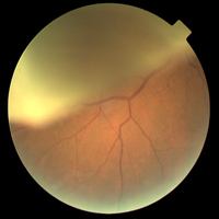
Retinal Detachment
The retina is multi-layered. Retinal detachment occurs when the neuroretina separates from the retinal pigment epithelium. Retinal detachment causes the retinal photoreceptor cells to be deprived of nutrient supply, damaging their visual function. Without proper treatment, the condition will cause permanent impairment to the vision and may progress to blindness.