 ENG
ENG
 繁中
繁中
 簡中
簡中
Our Facilities
Operation Theatre Equipment
(A) Cataract phacoemulsification system
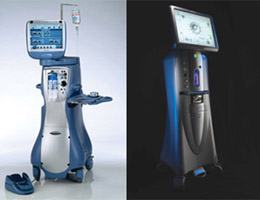
The cataract phacoemulsification system uses supermicroinvasive technology to melt and suck out the cataract piece, and then implant intraocular lens. The invasion is minimal and the surgical incision is ultra-fine. Our centre is equipped with an array of cataract phacoemulsification systems and a wide range of intraocular lenses of diverse functions to meet the specific needs of patients.
(B) High-speed retinal vitrectomy system
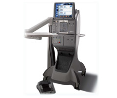
Our high-speed retinal vitrectomy system delivers an exceptional level of performance through state-of-the-art technology for the treatment of eye problems including retinal detachment.
(C) Cryosurgical system
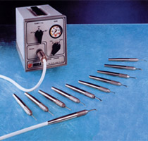
Our cryo surgical system is used to treat retinal detachment.
(D) Endoscopic dacryocystorhinostomy
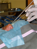
A minimal invasive surgical method to treat nasal lacrimal duct (tear pathway) obstruction. The advantages include “no scar” and a high success rate.
Laser Treatment System - Laser Therapy Machines
(1) 532 laser machine
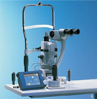
For the treatment of retinal perforation, glaucoma and maculopathy.
(2) YAG laser machine
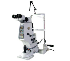
For the treatment of posterior capsular opacification after cataract surgery.
(3) PDT photodynamic laser therapy
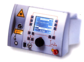
With intravenous injection of Visudyne, non-thermal laser is used to eliminate abnormal new blood vessel growth in the macula.
Examination and Diagnostic Equipment
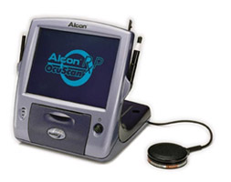
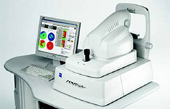
(1) Non-contact optical coherence biometry device
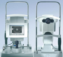
Through non-contact optical coherence biometric technology, we can measure the visual axis of the eye, depth of the anterior chamber, lens thickness and corneal refractive, etc. to obtain accurate data of the intraocular lens power in cataract cases, by which patients’ comfort and their after-treatment quality of vision are significantly enhanced.
(2) A-scan biometry
Ultrasonic contact technology measures the visual axis of more mature cataract patients for determination of the power of the intraocular lens to be implanted.
(3) B-scan biometry
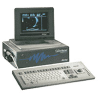
For the diagnosis of retinopathy.
(4) Fundus camera
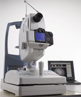
Fundus camera is used to take retinal photos.
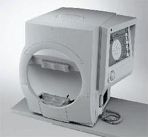
(5) Heidelberg confocal laser fundus imaging and spectral-domain optical coherence tomography scan joint-imaging system
With eye-tracking technology, the system performs a wider range of high-speed scan of the macula and retina; simultaneously, it generates eye fundus vessel and retinal tomography images. The system is highly helpful in diagnosing, monitoring and establishing the treatment for age-related macular degeneration, diabetic retinopathy and other retinal diseases and glaucoma.
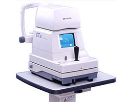
(6) Zeiss optical coherence tomography system
The system performs analyses of the retina, macula, optic nerve in glaucoma and optic disc, which help doctors diagnose, observe and treat glaucoma and maculopathy more effectively and accurately.
(7) Visual field analyzer
The device is used for the follow-up of glaucoma cases. It detects the visual field changes of glaucoma patients, providing guidelines for treatment.
(8) Non-contact tonometer
For the initial diagnosis of glaucoma.