 ENG
ENG
 繁中
繁中
 簡中
簡中
Age-related Macular Degeneration
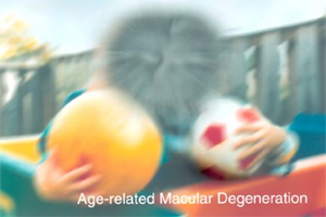
The macular contains a large number of photoreceptor cells to provide fine visual function and is located in the centre of the retina. Macular degeneration impairs the central vision, and if the condition is caused by aging, it is referred to as “age-related macular degeneration”. Age-related macular degeneration is one of the 3 major causes of blindness among people over the age of 55.
Symptoms
| . | Problem with colour discrimination. |
| . | Blurred central vision, especially obvious when reading or doing needle work. |
| . | Bent or deformed vision when looking at straight lines or distant scenes. |
| . | Gradually developing into darkness in the centre of vision, and a black hole will appear, forming a blind spot. |
Aged-related macular degeneration is classified into “dry” and “wet”. Most of the cases belong to the “dry macular degeneration”:
Dry Age-related Macular Degeneration: Yellow-white sediment is deposited in the retinal pigment epithelial cells of the macular, which weakens the photoreceptor cells, impairing the vision gradually. Although the patient will not lose their central vision completely in general, they will experience difficulties when doing fine visual activities such as reading, drawing and needle work, for which more light is required. Currently no effective medical approach is available to treat dry age-related macular degeneration.

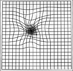
Wet Age-related Macular Degeneration: In wet age-related macular degeneration, abnormal neovascularization appears under the macular, which causes the macular to bleed and leak fluid, damaging the central vision. The visual deterioration is rapid yet early diagnosis and treatment can prevent vision from further damage. So if your vision becomes blurred or distorted, you should visit an ophthalmologist as soon as possible. Dry age-related macular regeneration can develop into the wet type. Wet age-related macular degeneration can cause severe vision deterioration within 3 months. If you have any doubts concerning this, especially if your vision suddenly deteriorates, you should go to see an ophthalmologist immediately without hesitation. The patient may use the” Amsler Grid” to monitor their own vision regularly at home. Concerns are indicated by the sight of any bent line or blurred image in the chart.
|
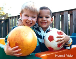
Normal Vision
|
Age-related Macular Degeneration
|
Amsler Grid Test
|
Causes of Age-related Macular Degeneration
The causes of dry and wet age-related macular degeneration are uncertain. Nevertheless, according to researches, the possible causes include genetics, dietary habits, smoking, diabetes, high blood pressure, glare and excessive emotional tension.
Prevention of Age-related Macular Degeneration
| . | Maintain a balanced diet and avoid high saturated fat and high cholesterol foods. |
| . | Vitamins A, C and E, and zinc and antioxidants can prevent and reduce the likelihood of age-related macular degeneration. Eat more green leafy vegetables and carotenoids, such as carrots, cereals, dried beans and fruits etc. |
| . | Eat less pan-fried, deep-fried and barbecued foods. |
| . | Quit smoking. |
| . | Wear UV protection sunglasses. |
The pre-treatment for age-related macular degeneration examination include: visual examination, fluorescent angiography and macular scan.
Treatment of Age-related Macular Degeneration
1. Photodynamic therapy After Verteporfin (Visudyne) is administered intravenously for 15 minutes, the doctor will apply a specific laser beam to activate the drug so that it will destroy and seal the abnormal blood vessels on the patient’s retina to reduce blood and fluid leakage. The purpose is to maintain the patient’s current vision or slow down their vision loss. The patient needs to receive 2 to 3 treatments in a year on average.
2. Intravitreal Injection Injecting anti-vascular endothelial growth factor (anti-VEGF) drugs into the eye can inhibit the forming and growth of new blood vessels, helping stabilize the condition of macular degeneration and slow down vision loss. However, in some cases, the vision continues to deteriorate after the administration. The result depends on the severity of the condition and individual patient’s reactions. Anti-VEGF intravitreal injections cannot revert permanent damage to the retina caused by age-related macular degeneration.
Avastin®(Bevacizumab) and Lucentis®(Ranibizumab) are two common anti-VEGF drugs.
| ·Lucentis®(Ranibizumab) | : FDA-approved medicine for the treatment of a variety of eye diseases. It is costly. |
| ·Bevacizumab(Avastin®) | : Not yet a registered eye medicine. It is less expensive. |
Results of a large-scale study comparing the efficacy of Avastin and Lucentis in treating wet age-related macular degeneration conducted by the U.S. National Institute of Health: the 1,208 patients surveyed who had been treated with either Avastin or Lucentis for 1 year had the same degree of vision recovery, which reveals that the 2 drugs exhibit no difference in efficacy. With regard to side effects, although both groups had cases involving serious adverse reactions, most of the cases were totally unrelated to the drugs’ recognized side effects.
Process of Intravitreal Injection
Intravitreal injection of anti-VEGF drugs is carried out after a disinfection process. Before injection, the patient’s pupil needs to be dilated, and local anesthesia and antibiotics are applied. During the procedure, the drug is administered into the vitreous to inhibit the forming and growth of new blood vessels, which will reduce fluid leakage and bleeding. After treatment, the patient needs regular follow-up to monitor the progress of the disease. According to many clinical studies, it is recommended that patients receive 3 consecutive injections in 3 months, and if new blood vessels appear, they have to undergo another treatment.
Disclaimer: The contents of this website are for reference only. They are not, and should not be used as, diagnoses, medical treatments or recommendations for any drug. For enquiries, please contact Champion Eye Centre.